Review from yesterday: diagram of bacterial cell (gram + and Gram -) differences
Optional parts
found in some bacteria:
Plasmids are extra bits of information and not required for cell functions.
Optional parts: Plasmids
*Small "extra" segments of DNA
* Generic
* Self replicating
* Easily passed
* (flash drive compared to hard drive)
r Plasmids = Resistance to antibiotics
* 3 Common Mechanisms of antibiotic resistance coded for on r plasmids:
- Efflux pump pumps out drug
- Enzymes break down drug
- Enzymes change drug
f plasmids = fertility genes
- initiate conjugation
- make sex pili
t plasmids = production of toxins
Common combined plasmid (picture example)
(one of the reasons diahrea is that it eases transmition of organism to others)
***draw plasmids in diagram***
**hairy stuff**
Sex Pili (Fig. 3.11)
Function: temporary (directions on F plasmid)
- tubular pilin protein, contracts
- connect cells: allow DNA transfer
*Do not limit to just one partner: picture of 1 s positive giving plasmid to three s negative cells.
Fimbrae (Fig. 3.10) made of pillin protein
f plasmids = fertility genes
- initiate conjugation
- make sex pili
t plasmids = production of toxins
Common combined plasmid (picture example)
(one of the reasons diahrea is that it eases transmition of organism to others)
***draw plasmids in diagram***
**hairy stuff**
Sex Pili (Fig. 3.11)
Appearance:
hollow tube that contracts down upon itself pulls them together. only contruct when there was another bacteria that did NOT have the plasmid. s positive, s negative... surface molecule that indicates that they do or don't have it..
- tubular pilin protein, contracts
- connect cells: allow DNA transfer
*Do not limit to just one partner: picture of 1 s positive giving plasmid to three s negative cells.
Fimbrae (Fig. 3.10) made of pillin protein
Appearance: short and numerous; proteinaceous
Function: for adhesion to surfaces
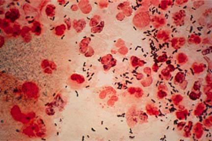
example: gonnorhia ... clean cervice (women).. urethra (men)
http://www.ncbi.nlm.nih.gov/pubmedhealth/PMH0004526/
example: gonnorhia ... clean cervice (women).. urethra (men)
http://www.ncbi.nlm.nih.gov/pubmedhealth/PMH0004526/
Appearance: hairlike structures
- Stiff, curved
- rotate
* counterclockwise = runs (positive simulus: food, good environment)
* clockwise = tumbles (negative such as toxin, bad environment)
- Stiff, curved
- rotate
* counterclockwise = runs (positive simulus: food, good environment)
* clockwise = tumbles (negative such as toxin, bad environment)
Function: movement
Fig. 3.9
Run: Flagellum moves in __counterclockwise_________
direction, bacterium moves ____quickly towards__________
Tumble: Flagellum moves in ___clockwise_____
direction, bacterium moves _____away__________
Taxis: (chemo, photo, etc.): movement in response to stimulus
* more runs, fewer tumbles if detect positive stimulus ahead
* Fewer runs more tumbles if detect negative stimulus ahead.
* more runs, fewer tumbles if detect positive stimulus ahead
* Fewer runs more tumbles if detect negative stimulus ahead.
Four
common arrangments:
- lophotrichous - tuft..... Lopho "tassle"
- monotrichous -One flagellan all by itself........ mono = "one"
- peritrichous -surrounding......... peri = all over exterior
- amphitrichous -both ends.........."amphi" = "both"
*** Gloey stuff**
Glycocalyx (Fig. 3.5)
- polysaccharide or glycoprotein
thick yellow, green, cloudy mucus = bacterial infection and not thin clear like your bodies natural response to a virus or allegries...
- slime layer = loose layer
- capsules = dens, thick layer
- biofilms = shared layer, many cells
Advantages of Glycocalyx/Capsules/Biofilms
- Confer increased pathogenicity, or help with survival:
1-neutralize drugs
2- fool/ delay immune response (capsule first)
3- adhere to surfaces
4- avoid phagocytosis (White blood cells eating of cell)
Help cell survive:
Help cell survive:
5- nutrient source during starvation
6- storage of toxic wast products
* dormant, non-reproductive structures
* formed inside cell in bad conditions
Sporulation process: cell creates an endospore
* layers of petidoglycan + protein = spore coat
* water removed
* Dipicolinic acid added- heat stablity
Spore coat composed of: petidoglycan
spores contain decreased amounts of:
spores contain high amounts of:
Highly resistant to harsh environmental
conditions:
- heat (survive boiling)
- harsh chemicals (alcohol, etc.)
- drying
- lack of nutrients
Germination in favorable conditions to normal vegetative cells.
·
Germination: spore to cell
Endospore structure and types:* Bacterical spores are used to help identify the species by using three characteristics:
- location
- size
- shape
*******oval large central endospore: green, pink.. force dye in spore? learn how to do it in lab*****

______________________________________________________________________________________________
*Review of drug targets discussed earlier Figure 10.2
- attack ribosome
- cell wall synthesis
- DNA or RNA synthesis
*ADDITIONAL DRUG TARGET-block enzyme
__________________________________________________________________________________________________
Unusual
Bacteria:
·
Archaea Fig. 3.26, 3.27
- ancient
- not "true" bacteria
- Cell wall has NO peptidoglycan (protein)
- Simpler than bacteria (have hooks and not a lot of other things that bacteria has)
- methanogens
- extremophiles
- thermophiles (hot temperatures)
- halophiles (salty environments)
- geothermal rift (archaea bacteria located)
·
Mycoplasma
Fig. 11.15
- Some pneumonias (e.g. "walking" pneumonia)
- no cell wall (can't survive if NOT in host tissue)
·
Rickettsia
and Chlamydia Fig. 21.1, 21.6
- Rocky Mountain Spotted Fever (Rickettsia) more come in NC then anywhere else in the world... rapid progressing disease...
- Chalmydia: eye or venereal disease
- replicate only inside host cell, burst out to infect new cells
- Spirochaetes Fig. 3.8
- Borrelia = lyme disease
- treponema = syphilis
- HUGE spiral cells
- axial filament of internal flagella for motility ( drills through tissue because the WHOLE cell turns not just the flagella to move)
Bacterial Growth
Bacteria Reproduce by Cloning...
- binary fission (Fig. 6.18, 11.2)= splitting in two.
- genome is copied.
- bidirectional
- topoisomerase fixes supercoils
- Cell elongates
- new cell wall and membrane separates the new cells.
- generation time= time between divisions
group activity?
* Ten bacterial cells were introduced into a test tube. In an hour, there are 80 cells present. What is the generation time of this particular species of bacteria?
10 >>> 20 >>> 40>>>80
divides 3 times in 60 minutes... 60 min/3 = 20 minutes
Generation time = every 20 minutes
Bacterial Growth Curve (Fig. 6.19, 6.21)
- Lag phase -
- Exponential growth phase -
- Maximum stationary phase -
- Exponential death phase -
- Minimum stationary phase OR crash
No comments:
Post a Comment